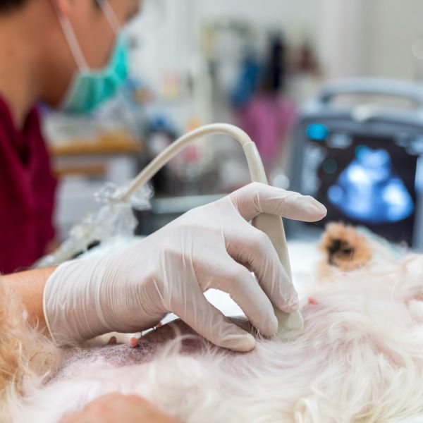
Pet Ultrasonography at South Broward Animal Hospital in Fort Lauderdale, FL
Unlock invaluable insights into your beloved pet’s health and well-being through the advanced ultrasonography services at South Broward Animal Hospital.
Understanding Ultrasonography: Probing the Depths of Pet Health
An ultrasound is a powerful diagnostic tool that creates a real-time image of an animal’s body. This composite reveals important information about internal processes including the circulatory, skeletal, and gastrointestinal systems, helping identify disease, blockages, and internal injury.
At South Broward Animal Hospital, our ultrasound services provide invaluable insights into your pet’s health. Utilizing advanced technology and experienced veterinary professionals, we conduct thorough real-time imaging of your pet’s internal organs. This non-invasive procedure allows us to detect a wide range of conditions, from gastrointestinal issues to cardiovascular abnormalities, with precision and accuracy. Whether assessing heart function, examining abdominal organs, or monitoring pregnancies, our ultrasound expertise ensures comprehensive care for your furry companion.
Unraveling Ultrasonography: How It Reveals Your Pet’s Health
An ultrasound works by broadcasting high-frequency sound waves that reflect off your pet’s internal structures. A small probe held against the skin collects the returning signals to create an image of the internal body, most commonly used to examine abdominal organs like the stomach, kidneys, liver, spleen, and gallbladder. An echocardiogram, or ultrasound of the heart, provides precise information about heart valves, blood flow, chamber size, and contractions. This tool is essential when assessing overall heart health and treating cardiovascular disease. Because an ultrasound doesn’t require radiation, it is also used to monitor pregnancies and fetal health in breeding pets.
When used in conjunction with other diagnostics tools like radiographs (X-rays), ultrasonography can detect a broad range of abnormalities including cardiovascular disease, skeletal fractures, some forms of cancer, soft tissue damage, foreign bodies, and organ disease. Completely painless and non-invasive, ultrasounds rank among the most precise diagnostic tools in the veterinary industry.
When Is Ultrasonography Necessary?
Ultrasonography may be recommended in various scenarios, including:
- Investigating gastrointestinal issues
- Evaluating urinary tract disorders
- Assessing cardiac function
- Monitoring pregnancy
- Detecting tumors or abnormal growths
Ultrasonography plays a crucial role in identifying abnormalities in organs like the liver, spleen, and pancreas, as well as in reproductive organs. It is also used to guide biopsies or aspirates, evaluate musculoskeletal injuries and abnormalities in soft tissues, and look for abnormalities or signs of disease in the thyroid gland, adrenal glands, and other endocrine organs.
During surgery, ultrasound guides vegetarians with precise imagery of internal structures. In wellness exams, it monitors the progression of chronic diseases or conditions over time, including assessing blood flow and circulation in specific organs or tissues and screening for congenital abnormalities or developmental issues.

